
Cell migration assay
Summary
Cell migration assays are used to compare mobility of cells under different conditions. It tests a cell’s ability to “crawl” across a solid substrate.
Also known as:
Scratch assay, wound-healing assay
Samples needed
A plate of confluent cells in a monolayer
Controls
The cell migration assay is nearly always used to compare mobility of cells +/- a treatment or intervention. Therefore, the usual mock treatment controls apply.
Method
First, cells of interest are grown to confluence in a monolayer. Then, a pipette tip or another object is used to create a scratch through the monolayer, leaving an area of a consistent width open and free of cells. Finally, the “scratch” or “wound” is monitored and photographed to determine if one treatment group refills the gap faster than another.
Interpretation
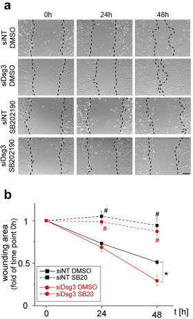 Figure 1. A cell migration assay in cells +/- siRNA for Dsg3 and +/- inhibitor of p38MAPK. Relevant section of caption for published figure reads: “(a) Representative bright-field images of HaCaT keratinocyte migration subjected to nontarget (siNT) or Dsg3 siRNA (siDSG3) and treated with solvent or with the p38MAPK inhibitor SB202190 (SB20). Bar is 150 mm. (b) Wound closure expressed as the remaining area uncovered by the cells. The scratch area at time point 0 hours was set to 1 (n = 4–6; *P < 0.05, #P< 0.05 vs. respective DMSO condition).” “Figure 3” by Vera Rötzer, Eva Hartlieb, Julia Winkler, Elias Walter, Angela Schlipp, Miklós Sardy, Volker Spindler, Jens Waschke[1]. [Image description]
These microscopy images show that cells with decreased Dsg3 expression “heal the wound” faster by migrating to fill the scratched area. Therefore, the reader can conclude that one normal function of Dsg3 is to suppress cell migration. However, regardless of whether or not Dsg3 is expressed, if p38MAPK is inhibited, wound closure will not occur. Therefore, the migration-suppressing function of Dsg3 is dependent on p38MAPK activity.
Image Descriptions
Figure 1 image description: Data from a cell migration assay. Upper row shows cells from Dsg3+/+ mice and lower shows cells from Dsg3-/- mice. The columns show 0 h, 24 h, and 48 h. [Return to Figure 1]
- Rötzer, V., E. Hartlieb, J. Winkler, E. Walter, A. Schlipp, M. Sardy, V. Spindler, and J. Waschke. 2016. Desmoglein 3-dependent signaling regulates keratinocyte migration and wound healing. Journal of Investigative Dermatology 136:301-310. ↵
Touching, completely covering the plastic bottom of a culture vessel
Single layer, not growing on top of one another
