
Soft agar colony formation assay
Summary
The soft agar colony formation assay is a test to determine whether cells are anchorage-independent. Anchorage independence is a characteristic of cells that have undergone malignant transformation.
Also known as: Test of anchorage independence
Samples needed: Cells capable of growing in tissue culture
Controls:
The soft agar colony formation assay is nearly always used to compare anchorage-independence of cells +/- a treatment or intervention. Therefore, the usual mock treatment controls apply.
Method:
Normal, non-transformed cells only grow attached to a solid substrate. This characteristic is called anchorage dependence. A distinctive feature of transformed cells is their ability to grow without that solid substrate; they are anchorage independent. To perform the test for anchorage independence, cells are plated in agar, a soft, jelly-like substance, and supplied with the necessary nutrients and growth factors. After 2-3 weeks, the plates are stained to aid in colony identification, then photographed, and the colonies are counted. [1]
Interpretation:
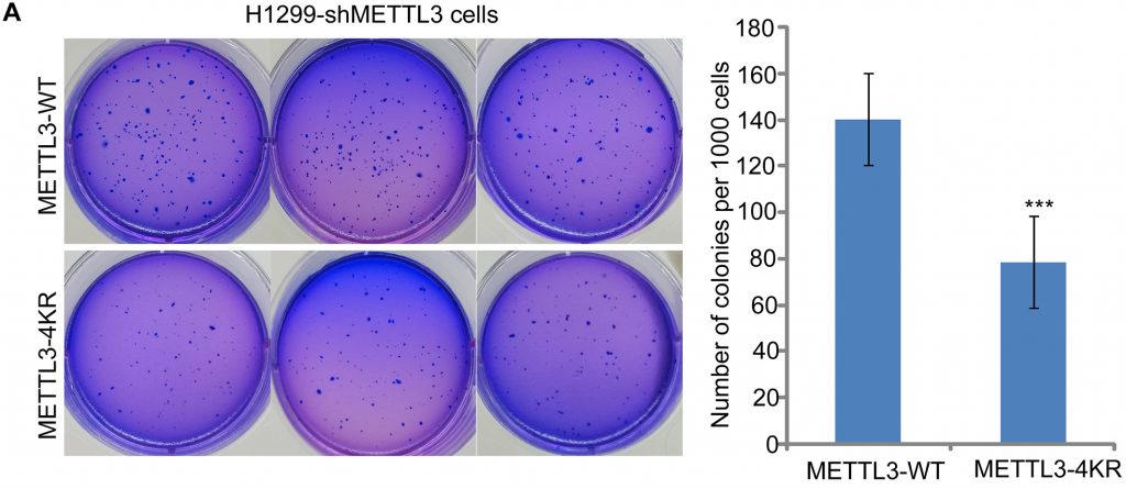 Figure 1. A soft agar colony formation assay on H1299 cells with either wild type METTL3 protein or METTL3 with a mutation that turns four key lysine residues to arginine residues to prevent SUMOylation. Relevant section of caption for published figure reads: “(A) The mutation of 4KR in METTL3 reduces the soft-agar colony formation of H1299 cells. H1299-shMETTL3 cells stably re-expressed METTL3-WT or METTL3-4KR cell lines were seeded in 2 ml of medium containing 10% FBS with 0.35% soft agarose at 1000 per well and layered onto 0.6% solidified agarose. The photographs were taken 14 days after seeding, and the number of colonies were counted and analysed. Each value represents the mean±s.e.m. of three independent experiments with triplicates.” “Figure 5” by Yuzhang Du, Guofang Hou, Hailong Zhang, Jinzhuo Dou, Jianfeng He, Yanming Guo, Lian Li, Ran Chen, Yanli Wang, Rong Deng, Jian Huang, Bin Jiang, Ming Xu, Jinke Cheng, Guo-Qiang Chen, Xian Zhao, Jianxiu Yu[2]. [Image description]
This figure shows the reader that in H1299 non-small cell lung cancer(NSCLC) cells, the SUMOylation of METTL3 is important for the process of transformation. This is evident because if METTL3 cannot be SUMOylated (4KR mutatnt), significantly fewer colonies are able to form in soft agar.
Image Descriptions
Figure 1 image description: Left: Six cell culture plates containing purple-stained soft agar. In each plate, there are some darker purple spots showing cell colonies. There are more colonies in the three wild type plates compared to the mutant plates. Right: A column graph quantifying colony growth. The wild type condition shows about 140 colonies per 1000 cells, whereas the mutant condition shows about 80. The difference is significant. [Return to Figure 1]
- Borowicz, S., M. Van Scoyk, S. Avasarala, K. Karuppusamy Rathinam Manoj, J. Tauler, K. Bikkavilli Rama, and R. A. Winn. 2014. The soft agar colony formation assay. JoVE :e51998. ↵
- Du, Y., G. Hou, H. Zhang, J. Dou, J. He, Y. Guo, L. Li, R. Chen, Y. Wang, R. Deng, J. Huang, B. Jiang, M. Xu, J. Cheng, G. Chen, X. Zhao, and J. Yu. 2018. SUMOylation of the m6A-RNA methyltransferase METTL3 modulates its function. Nucleic Acids Research 46:5195-5208. ↵
Gaining the properties of a cancer cell
