
Immunofluorescence microscopy
Summary
Immunofluorescence microscopy shows where in a cell a particular protein is located.
Also known as:
Immunofluorescence, IF
Samples needed
Fixed cells or tissue on a slide
Controls
Frequently, in addition to antibodies for the protein of interest, cells are also stained with DAPI. DAPI binds to DNA, thus marking the cell nucleus. When DNA-bound, DAPI fluoresces, emitting blue light.
Often times, other controls for IF are not shown in a published work. However, if a researcher is using new IF antibodies for the first time, several controls are useful. A positive control that contains the protein of interest tells the researcher if the antibody works well in IF and/or if the conditions used for staining are optimal. A control without the primary antibody ensures that the secondary antibody is binding specifically to the primary antibody and not other proteins. Finally, a control without the secondary antibody ensures that any observed signal is not autofluorescence of the cells or tissues.
Method
To stain a sample for immunofluorescence microscopy, samples are first fixed, then solubilized. Solubilizing creates holes in the plasma membrane to allow the antibodies to enter the sample. The sample is then blocked, often with milk protein, to discourage any non-specific binding of antibodies. Next, the sample is incubated in a primary antibody, which specifically binds to the protein of interest. Then, the sample is incubated in a secondary antibody, which binds to the primary antibody based on what species the primary antibody was raised in. (For instance, the secondary antibody might be something like anti-rabbit IgG.) The secondary antibody is chemically attached to a fluorescent molecule that allows detection with a fluorescence microscope.
Interpretation
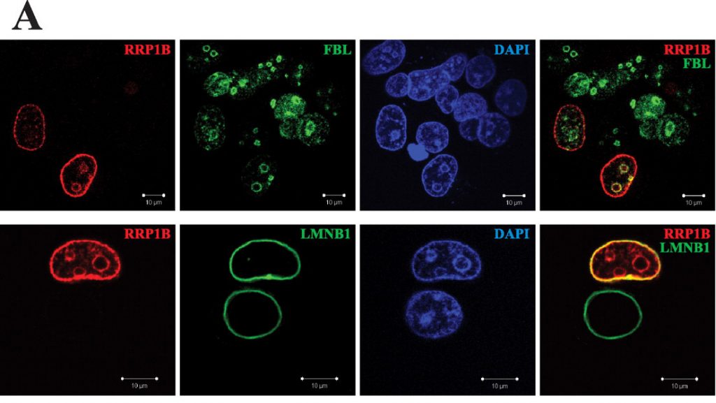 Figure 1. Two immunofluorescence experiments assessing intracellular localization of protein RRP1B. Relevant section of caption for published figure reads: “A, co-IF followed by confocal microscopy was performed to define the cellular localization of RRP1B. Hemagglutinin-tagged, full-length RRP1B was ectopically expressed in HeLa cells to define its cellular localization. Cells were co-stained for ectopic RRP1B (red), either the nucleolar marker fibrillarin (FBL; top) or the nuclear envelope marker lamin B1 (LMNB1; bottom) (green), and double-stranded DNA (DAPI; blue). ” “Figure 2” by Nigel P.S. Crawford, Hailiu Yang, Katherine R. Mattaini, Kent W. Hunter[1]. [Image description]
The reader can tell that these images focus on cell nuclei, because of the blue rounded structures in the DAPI channel. The authors are interested in the protein RRP1B and want to determine where it is within the cell. In the top panels, cells have been stained for both RRP1B and the protein fibrillarin, which is known to localize to the nucleolus. The top right panel is the overlay of the red (RRP1B) and green (fibrillarin) channels. In the image, locations with both red and green signals appear yellow, indicating that both proteins are found in that location. This is one piece of evidence that RRP1B is found in the perinucleolar region (region surrounding the nucleolus). The bottom panels are similar, but the green channel shows lamin B1, a protein that is known to localize to the nuclear envelope. The bottom right panel shows a nucleus with a yellow outline, demonstrating that RRP1B is also found at the periphery of the nucleus.
Note that in this experiment RRP1B was expressed ectopically, and the primary antibody detects the HA-tag on RRP1B, not RRP1B itself. Therefore, not all cells show the presence of HA-RRP1B. RRP1B may be present in other cells, but since it is the endogenous protein and not the tagged protein, it is not detected in this experiment. This is not considered ideal, but if an immunofluorescence antibody for the protein of interest is not available, this is one option to determine intracellular localization.
Image Descriptions
Figure 1 image description: Two immunofluorescence experiments with four panels each. In both experiments, RRP1B (red) outlines the nucleus and nucleoli; DAPI (blue) stains throughout the nucleus. In the first experiment, FBL (green) marks the nucleoli; in the red/green overlay, the nucleoli are outlined in yellow. In the second experiment, LMNB1 (green) outlines the nucleus; in the red/green overlay, the nucleus is outlined in yellow. [Return to Figure 1]
- Crawford, N. P. S., H. Yang, K. R. Mattaini, and K. W. Hunter. 2009. The metastasis efficiency modifier ribosomal RNA processing 1 homolog B (RRP1B) is a chromatin-associated factor. Journal of Biological Chemistry 284:28660-28673. ↵
Proteins are one class of biological macromolecules. They are made of amino acid building blocks and have many biological functions including catalysis, structure, transport, signaling, and others.
Preserved in a way that stops all reactions and increases the stability of a sample; fixed samples are no longer living
A protein that binds very specifically to a protein of interest, normally produced by an organism's adaptive immune system to protect against pathogens, but also used in a variety of biotechnological applications
4′,6-diamidino-2-phenylindole, used to label DNA (and therefore the nucleus) in fluorescence microscopy
Naturally occurring fluorescence from cells or tissues that is not due to specific binding of a fluorescently labeled antibody
Expressed from a plasmid transfected into a cell rather than from the endogenous gene
A nine amino acid sequence from the influenza hemagglutinin protein; if a protein is expressed from a genetically-modified gene, the HA-tag can be added to make detection easier
