
Transwell migration and invasion assay
Summary
Transwell migration and invasion assays are used to determine if cells can move through a porous membrane (migration assay) or both the membrane and an extracellular matrix (invasion assay). Invasiveness is a characteristic feature of more advanced transformed cells, as it is necessary for metastasis.
Also known as:
Boyden chamber assay, modified Boyden chamber assay
Samples needed
A plate of confluent cells in a monolayer
Controls
The transwell migration and invasion assays are nearly always used to compare mobility and/or invasiveness of cells +/- a treatment or intervention. Therefore, the usual mock treatment controls apply.
Method
A chemoattractant is placed in the lower chamber of a cell culture plate. The cells are placed on top of a porous transwell membrane alone for the mobility assay or one covered with extracellular matrix for the invasion assay. Following an incubation period that is specific to the cell type, cells that have crossed the membrane will be stained and counted[1].
Interpretation
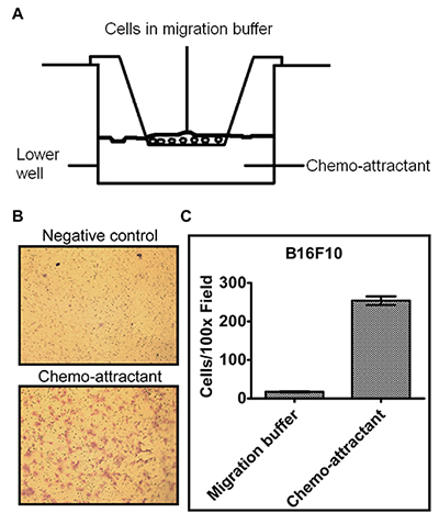
Figure 1. A schematic of the setup for a transwell assay and an example of representative results. Relevant section of caption for published figure reads: “(A) A diagram of the transwell insert apparatus used to measure cell migration and invasion. (B) Representative pictures of B16F10 cell transwell migration. Cell migration buffer and NIH3T3 cell conditioned medium were added to the lower chamber as the negative control and chemo-attractant, respectively. After cell migration and staining with crystal violet as described in the Procedure, pictures of the migrated cells (purple stained) were taken using a microscope with a 10x objective (total magnification 100x). Pores of the membranes could also be observed as the numerous small, round and dark colored dots in the picture. (C) Quantification of cells migrating toward the migration buffer or chemo-attractant(Average of 5 picture fields at 100x total magnification).” “Figure 3” by Calvin R. Justus, Nancy Leffler, Maria Ruiz-Echevarria, and Li V. Yang[2]. [Image description]
This figure shows that B16F10 melanoma cells only cross the transwell membrane when the liquid in the lower chamber is cell medium that has been “conditioned” by NIH3T3 cells. (NIH3T3 are immortalized mouse embryonic fibroblasts.) This means that in a different cell culture dish, NIH3T3 cells were grown in cell culture medium, then the medium only (not the cells) were placed in the lower chamber for the transwell assay. This “conditioned” medium will now contain any proteins and chemicals secreted by the NIH3T3 cells. The results show that the NIH3T3 conditioned medium contains a chemoattractant that promotes migration of the melanoma cells across the transwell membrane. Note that because there is no extracellular matrix used to coat the transwell insert, this is considered a migration assay and not an invasion assay.
Image Descriptions
Figure 1 image description: Schematic and example data from a transwell migration assay. Panel A shows the schematic. The lower chamber is about halfway full of liquid containing a chemoattractant. The upper chamber rests on top, with the bottom of the upper chamber submerged in the liquid in the lower chamber. The cells are in the upper chamber. Panel B shows two images of cell stains. The image with chemoattractant shows more cells that that without. Panel C is a column graph showing between 200-300 cells per 100x field with chemoattractant and almost none without. [Return to Figure 1]
Spread of cancer cells from the site of the primary tumor into other tissues
Touching, completely covering the plastic bottom of a culture vessel
Single layer, not growing on top of one another
Chemical that attracts cells
Capable of dividing indefinitely in cell culture
